Research News
Super-resolution microscope allows visualization of the mechanism that maintains cell polarity —The key is to repeatedly establish temporary polarity—
Cells are not uniform spheres; they generally come in a variety of disparate shapes. In the broadest sense, this variation in shapes is known as cell polarity, and it is an essential property for a variety of cell functions. Growth in accordance with their polarity allows cells to shape themselves in forms appropriate to their function. It has been found that the establishment and maintenance of polarity is governed by the interdependent relationship between the polarity marker protein on the plasma membrane (cell membrane), actin, the microtubule cytoskeleton, and membrane vesicle transport. The polarity marker determines the polarity site, and with membrane vesicle transport toward it, site-specific growth (polarity growth) is achieved. However, when the plasma membrane elongates due to fusion of membrane vesicles, there have been questions about how polarity markers are maintained without being scattered over the elongated plasma membrane.Cells are not uniform spheres; they generally come in a variety of disparate shapes. In the broadest sense, this variation in shapes is known as cell polarity, and it is an essential property for a variety of cell functions. Growth in accordance with their polarity allows cells to shape themselves in forms appropriate to their function. It has been found that the establishment and maintenance of polarity is governed by the interdependent relationship between the polarity marker protein on the plasma membrane (cell membrane), actin, the microtubule cytoskeleton, and membrane vesicle transport. The polarity marker determines the polarity site, and with membrane vesicle transport toward it, site-specific growth (polarity growth) is achieved. However, when the plasma membrane elongates due to fusion of membrane vesicles, there have been questions about how polarity markers are maintained without being scattered over the elongated plasma membrane.
A research group lead by University of Tsukuba Faculty of Life and Environmental Sciences International Tenure Track Assistant Professor Norio Takeshita (who holds a concurrent post as Group Leader of the Karlsruhe Institute of Technology Department of Applied Microbiology) has succeeded in using a super-resolution microscope to visualize the mechanism by which cell polarity is maintained.
Professor Takeshita's study used a super-resolution microscope to clarify the behavior of polarity markers that could not be visualized with conventional fluorescence microscopes. The study employed a fluorescence microscope with super-resolution capability to image a fungal model with a filamentous shape and visualize the behavior of polarity markers, clarifying their relationship to exocytosis (the fusion of a membrane vesicle to a plasma membrane) and microtubules.
Original Paper
Yuji Ishitsuka, Natasha Savage, Yiming Li, Anna Bergs, Nathalie Grün, Daria Kohler, Rebecca Donnelly, G. Ulrich Nienhaus, Reinhard Fischer, Norio Takeshita, Superresolution microscopy reveals a dynamic picture of cell polarity maintenance during directional growth, Science Advances 1(10),DOI: 10.1126/sciadv.1500947(2015)
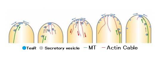
Transient polarity model