Research News
MRI Observation of Human Embryos at a One-Hundredth-of-Millimeter Resolution
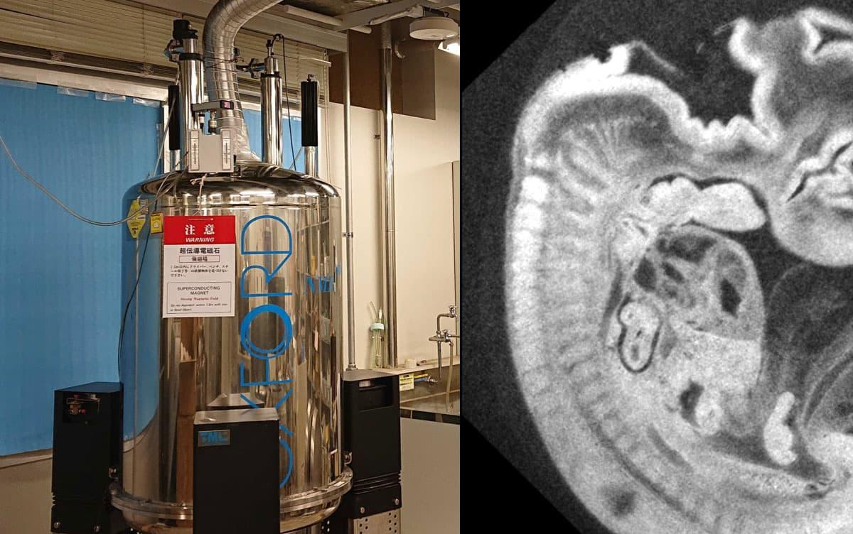
Researchers from the University of Tsukuba have improved the imaging resolution of the magnetic resonance (MR) microscope, which is used to obtain high-resolution images of the interiors of small objects. This improvement has achieved a spatial resolution of one-hundredth of a millimeter, which offers remarkably higher clarity--several tens of times better than previous versions. Consequently, one can obtain the microscopic structures of human embryos, particularly those of the brain, central nervous system, and other organs. Such technological advancements are poised to substantially impact human embryology.
Tsukuba, Japan—Magnetic resonance (MR) microscope has previously been used to observe chemically fixed specimens of human embryos during their early developmental stages. This tool has been instrumental in advanced studies of human embryology. MR microscopes enable researchers to visualize the development and growth of organs throughout various stages in three dimensions, leading to the creation of detailed three-dimensional morphological models. However, conventional MR microscopes are limited by their maximum spatial resolution of approximately four-hundredths of a millimeter, which occasionally results in blurred or lost small structures within human embryos.
To address this limitation, the research team has introduced a high-resolution MR microscope capable of a spatial resolution of one-hundredth of a millimeter. This leap in resolution clarity surpasses conventional capabilities, facilitating intricate detailing of the brain, central nervous system, and other organs in human embryos. This was achieved via enhancements in hardware, imaging methods, and the integration of efficient data collection and reconstruction techniques, such as compressed sensing technology.
In magnetic resonance imaging (MRI), there is no direct correlation between the set pixel size and the actual resolution; various factors resulted in reducing the resolution of the images. However, after testing the resolution using well-defined artificial structures designed for image quality evaluation, the research team found a match between the set pixel size and the achieved resolution. Moreover, upon comparing this MRI image with an optical microscope image of a human embryo specimen from the same developmental stage, results showed that the microstructure was precisely captured.
Such groundbreaking results emphasize that this improved resolution effectively captures the microstructure of the human embryo. This technique promises to revolutionize human embryology research, enabling the creation of a high-resolution atlas of brain and other organs.
###
This work was supported by JSPS KAKENHI Grant Number JP21H01333.
Original Paper
- Title of original paper:
- High-resolution MRI for human embryos with isotropic 10 µm resolution at 9.4 T
- Journal:
- Journal of Magnetic Resonance
- DOI:
- 10.1016/j.jmr.2023.107545
Correspondence
Associate Professor TERADA Yasuhiko
Institute of Pure and Applied Science, University of Tsukuba
Related Link
Institute of Pure and Applied Sciences
