Research News
Advanced Assistive Technology for Predicting Organ Deformation During Radiotherapy Utilizing Image Information
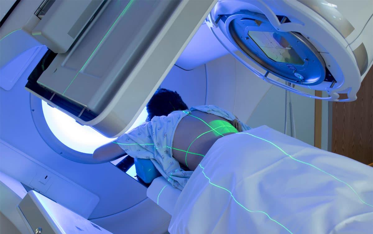
 Image by Mark_Kostich/Shutterstock
Accurate prediction of organ deformation during radiation therapy is pivotal for enhancing the treatment efficacy. Researchers from the University of Tsukuba have developed a technology capable of capturing real-time cross-sectional images of the affected area, thereby computing the three-dimensional motion of organs from these images. Consequently, this technology can precisely predict the deformation of the pancreas and other organs based on their position with respect to the surrounding organs.
Tsukuba, Japan—Radiation therapy, employed for treating cancer and other ailments, is distinctive for its minimally invasive nature, facilitating outpatient treatment and a fast return to society. However, a notable challenge arises due to the potential impact of radiation on the adjacent healthy organs, especially while applying high radiation doses to diseased tissues that are in motion. Regular movements such as breathing can be easily predicted; however, irregular movements initiated by contact with the surrounding organs are difficult to predict. In this study, a novel technique was devised to predict the three-dimensional motion of each organ based on its position with respect to the surrounding organs. This was achieved by acquiring cross-sectional (two-dimensional) images from three different orientations of the affected area in real time during radiation therapy. Additionally, a cross-section selection method was formulated to choose the most accurate cross-sectional image for analysis.
To validate this technique, the researchers assessed the position of the pancreas using publicly available MRI data of 20 cases. The results showed that the pancreas could be located with an error of 5.11 mm when only one directional cross-sectional information was utilized, which markedly reduced to an error of 2.13 mm when all three directions were employed. Furthermore, in some instances, the accuracy of the results matched with the ideal scenario wherein the three-dimensional information had been obtained in advance.
The findings of this research hold promising implications for practical applications, potentially paving the way for radiation therapy protocols that minimize radiation exposure to surrounding healthy organs, thereby promoting safer radiation therapy.
###
This work was partly supported by AMED under Grant Number by JP23YM0126803. (Corresponding author: Yoshihiro Kuroda)
Original Paper
- Title of original paper:
- 2D Slice-driven Physics-based 3D Motion Estimation Framework for Pancreatic Radiotherapy
- Journal:
- IEEE Transactions on Radiation and Plasma Medical Sciences
- DOI:
- 10.1109/TRPMS.2023.3313132
Correspondence
Professor KURODA Yoshihiro
Institute of Systems and Information Engineering, University of Tsukuba
Related Link
Institute of Systems and Information Engineering (in Japanese)


