Research News
Selective Destruction of Cancer Cells through Autophagy Mechanism: Innovations in Photodynamic Therapy Using Polphylipoprotein
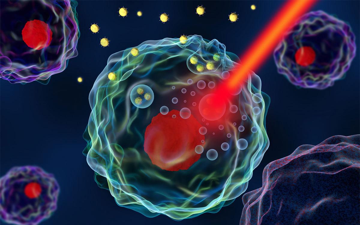
 Image by Meletios Verras/Shutterstock
Researchers at the University of Tsukuba observed the effects of photodynamic therapy (PDT) using a novel photosensitive substance, polphylipoprotein (PLP), on both normal and cancer cells. Results demonstrate that PLP selectively destroys cancer cells via the autophagy mechanism, marking a groundbreaking advancement in effective cancer therapy.
Tsukuba, Japan—In Japan, cancer has been the leading cause of death since 1981. At present, surgery, chemotherapy, and radiotherapy are the main treatment options. However, these treatments considerably reduce postoperative quality of life; hence, the development of new treatment methods is highly desired. Photodynamic therapy (PDT) is a new treatment method that is gaining attention owing to its lesser invasive nature than other treatment methods. In PDT, a photosensitizer is first injected into the body. Then, the diseased area is exposed to light, inducing a photochemical reaction in the photosensitizer in the region. This generates reactive oxygen species (ROS), resulting in the destruction of the cancer cells. To improve the performance of PDT, a deep understanding of the mechanism by which light-sensitive substances specifically accumulate in cells and how ROS efficiently kills only cancer cells is crucial.
Researchers highlighted a recently developed photosensitizer, polphylipoprotein (PLP), known for its high therapeutic efficacy. However, the underlying mechanisms of PLP's effectiveness remained unclear.
Using confocal superresolution microscopy, the research team investigated the effectiveness of PDT with PLP on two cell lines: the normal rat gastric epithelial cell line RGM1 and a gastric mucosa-derived cancer-like mutant RGK1 (a cancer cell of the same origin). The results revealed that PLP accumulates in the membranes of intracellular vesicles, known as phagosomes. These are produced early in the autophagy mechanism, which degrades and reuses foreign substances and proteins in the cell. The autophagy mechanism proceeds when cancer cells, which are rapidly proliferating, are starved. Post-PDT application, the phagosome membranes in RGK1 cells ruptured, releasing hydrolytic enzymes and ROS along with the materials produced from the breakdown of their own cytoplasmic components such as proteins, lipids and cellular organelles such as mitochondria and endoplasmic reticulum. These substances diffused into the cell, thereby leading to cell necrosis. In contrast, in RGM1, small phagosomes coalesced to form large phagosomes without rupture.
This phenomenon elucidates a unique and novel mechanism by which PLP selectively induces necrosis in cancer cells by exploiting the autophagy mechanism under starvation and using their degraded contents. This discovery opens new avenues for cancer therapy development, including the high selectivity and potential efficacy of PLP-PDT.
###
This work was supported by Grant-in-Aid for Scientific Research (23H00264) from Japan Society for the Promotion of Science and Japan Core Research Evolutional Science and Technology (CREST) (JPMJCR1875).
Original Paper
- Title of original paper:
- Polphylipoprotein-induced autophagy mechanism with high performance in photodynamic therapy
- Journal:
- Communications biology
- DOI:
- 10.1038/s42003-023-05598-0
Correspondence
Professor SHIGEKAWA Hidemi
Department of Applied Physics, Institute of Pure and Applied Sciences, University of Tsukuba / R&D Center for Innovative Material Characterization
Visiting Associate Professor TANIKAWA Atsushi
Institute of Pure and Applied Sciences, University of Tsukuba / Manager, TAKANO Co. LTD
Related Link
Institute of Pure and Applied Sciences
R&D Center for Innovative Material Characterization


