


Research News
New Insights into Neuron-Microglia Interactions During Brain Aging
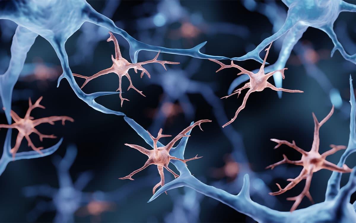 Image by ART-ur/Shutterstock
Researchers at University of Tsukuba identified small nuclear fragments known as micronuclei while studying neurons in aged mice. They demonstrated that microglia—the brain's resident immune cells—take up these micronuclei, leading to changes in microglial morphology and gene expression associated with inflammation and vascular function.
Tsukuba, Japan—As mammals age, their brains show chronic inflammatory responses and a gradual decline in vascular function. Microglia, the resident immune cells of the brain and spinal cord, play a central role in regulating brain aging. Recent studies have shown that microglia undergo profound changes in their properties and functions throughout life—from embryonic development to aging—in response to their surrounding environment. The research team has also reported that neuron-derived micronuclei (small extranuclear structures distinct from the primary nucleus) contribute to changes in microglia during development (Yano S et al., Nat Neurosci 2025). However, it remained unclear whether neuronal micronuclei also influence microglial function during aging.
To investigate whether micronuclei influence microglial function during aging, the research team analyzed the brains of aged mice in which neuronal nuclear membranes were labeled with a fluorescent protein (GFP). The analysis revealed a significant increase in micronuclei in aged mice compared with young mice. This increase was particularly evident in the deeper layers of the cerebral cortex. The micronuclei also contained markedly less DNA than micronuclei observed in young mice. Transmission electron microscopy revealed that micronuclei formation was closely associated with infolding (invagination) of the neuronal nuclear envelope. Using in vivo live imaging with two-photon excitation microscopy, the researchers visualized this process in real time.
The researchers observed that microglia internalized the micronuclei, leading to changes in both microglial morphology and gene expression. Specifically, microglia that had internalized micronuclei showed increased expression of gene clusters involved in heparan sulfate biosynthesis. Heparan sulfate proteoglycans—core proteins modified with heparan sulfate, a type of glycosaminoglycan—play an important role in regulating inflammatory responses and vascular function. Overall, these findings provide new insights into how neurons and microglia communicate and may help clarify the molecular basis of age-associated inflammation and vascular dysfunction in the brain.
###
This work was supported by the Gout and uric acid foundation (FT), Sumitomo foundation (FT), Ono Pharmaceutical foundation for Oncology, Immunology, and Neurology (FT, YK), AMED-PRIME [ADI07313 (FT)], JSPS Research Fellowship for Young Scientists [25KJ0660 (CM)], and partly supported by Center for Quantum and Information Life Sciences, University of Tsukuba (FT). The in vivo imaging was supported by the Cooperative Study Program of National Institute for Physiological Sciences [25NIPS301(FT)] and JSPS KAKENHI Grant Number JP22H04926(CM), Grant-in-Aid for Transformative Research Areas — Platforms for Advanced Technologies and Research Resources "Advanced Bioimaging Support."
Original Paper
- Title of original paper:
- Age-associated neuronal micronuclei formation and transfer to microglia
- Journal:
- Frontiers in Aging Neuroscience
- DOI:
- 10.3389/fnagi.2026.1787252
Correspondence
MAEDA Chihiro
Doctoral Program in Biology, Degree Programs in Life and Earth Sciences, Graduate School of Science and Technology, University of Tsukuba
Assistant Professor TSURUTA Fuminori
Institute of Life and Environmental Sciences, University of Tsukuba
Related Link
Institute of Life and Environmental Sciences

