TSUKUBA FRONTIER
#037 Connecting Structural Analysis of Proteins to Drug Discovery: Uncovering the Structures of Biomolecules with Cryo-Electron Microscopy
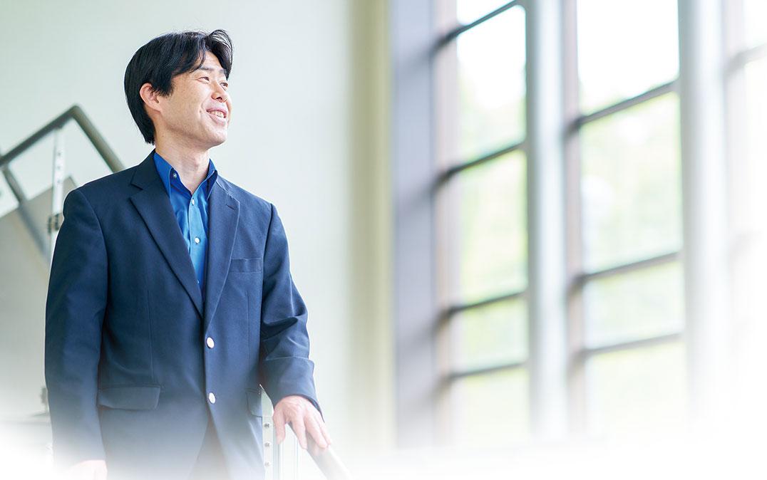
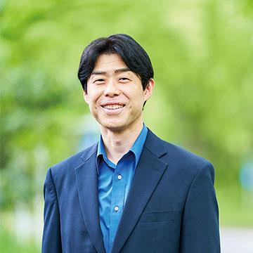
Professor IWASAKI Kenji, Life Science Center for Survival Dynamics, Tsukuba Advanced Research Alliance

Proteins are one of the building blocks of our bodies. They each have unique functions that interact in intricate ways to carry out activities that support life, and are composed of just 20 types of amino acids. The arrangement and spatial structure of amino acids within proteins are what produce their diverse range of functions, and defects in the amino acid structure can cause disease. Prof. Iwasaki leverages state-of-the-art cryo-electron microscopy to analyze previously unobservable 3D structures of proteins in order to find clues about the mechanisms underlying their functions.
The challenge of analyzing biomolecules

Microscopes are commonly used to observe the detailed structure of substances. Optical microscopes magnify images by shining visible light on the surface of a sample, and electron microscopes use electron beams instead of light to observe samples at higher magnifications. To prevent disturbance of electrons by surrounding gas molecules, the inside of the electron microscope is a vacuum. However, this makes it tricky to observe biomolecules with an electron microscope. Water in biomolecules evaporates rapidly in a vacuum, which alters the properties of the sample. Even if the water is frozen, electrons will scatter in the ice crystals and be unable to reach the sample. Therefore, it was long considered impossible to observe proteins and other biomolecules with an electron microscope.
That is why X-ray crystallography has been used instead. With this method, the structure of a molecule is determined by scattering X-rays off a crystal, but this requires that the sample be crystallized. Although proteins can also be crystallized, this process is challenging and presents a high hurdle to overcome when preparing a sample for observation.
For this reason, despite the great need for structural analysis of biomolecules as a way to understand biological phenomena and discover new drugs, the difficulty of finding a decisive analytical method has continued to frustrate researchers.
Cryo-electron microscopy opens up new possibilities
Then cryo-electron microscopes came on the scene. "Cryo-" means "to freeze" or "cold temperature." The basic structure of a cryo-electron microscope is the same as a conventional electron microscope, but as the name implies, the inside of the instrument is chilled using liquid nitrogen. A sample frozen in a near-liquid state is inserted into this chilled instrument. Cooling the sample also prevents damage to the sample from the electron beam. This innovation has suddenly opened up the possibility of using electron microscopy for structural analysis of biomolecules.
The idea may seem simple at first glance, but this technology won the Nobel Prize in Chemistry in 2017. This is because structural analysis of biomolecules was such an important challenge. Advances in camera and analytical technology were particularly groundbreaking. Modern structural analysis requires more than just high performance from the equipment itself; it is only possible with a camera capable of capturing precise images and an algorithm to process the data and construct a 3D image. For this reason, cloud technology and powerful computers that can handle vast amounts of video data are also essential. These technologies were developed and combined at just the right time for the cryo-electron microscope to be used to its full advantage.
Cryo-electron microscopy is also being used for structural analysis of the coronavirus (SARS-CoV-2) spike protein. Because the structure of the spike's tip changes after infection, an antibody drug that keeps the spike protein in its pre-infection state would be an effective measure against COVID-19. The superior performance and user-friendliness of cryo-electron microscopes made it possible to conduct this kind of structural analysis in a timely manner early in the pandemic.
Searching for drugs to treat a rare disease
Prof. Iwasaki's current focus is developing drugs for a rare cancer called synovial sarcoma. He became motivated when a family member developed this disease. After hearing the news, he immediately asked the treating physician to conduct joint research and provide genetic data, which Prof. Iwasaki then began to analyze. Although synovial sarcoma is known to be caused by the fusion of two proteins, the mechanism leading to its onset is still a mystery. One hypothesis is that onset is triggered when this strange fusion protein expels the normal protein that is naturally produced in the body. Prof. Iwasaki successfully uncovered a part of this process through structural analysis. Precise analysis using cryo-electron microscopy also played a decisive role in this achievement. Prof. Iwasaki believes that approaching research from the perspective of his own specialty field of structural analysis rather than from conventional methods used in the medical world, should generate multifaceted findings about diseases. This philosophy has proved effective, and Prof. Iwasaki feels he is very close to finding a clue to a cure.
Large pharmaceutical companies often cannot take on such research on rare diseases, so it should be tackled in an academic setting. Although this is quite a challenging topic to research, being at the University of Tsukuba offers Prof. Iwasaki the great advantage of an environment that facilitates collaboration. With the help of various collaborators, he has been able to achieve better results than he imagined in a relatively short period of time.
An unpopular field jumps to the cutting edge
When Prof. Iwasaki was a graduate student, he studied viruses and was amazed at how quickly researchers around him were conducting experiments. Thinking that he had no chance to succeed as a virus researcher, he decided to slightly shift his field of study a bit, and received advice from a professor to try electron microscopy research. He plunged right into the work without knowing anything. This kind of research requires persistent effort, as it can take years to collect data and determine a single structure, but Prof. Iwasaki thought that he might have a good chance at success because so few people specialized in the field.
However, X-ray crystallography was still the mainstream method used for structural analysis at that time. Electron microscopy was so unpopular due to its poor resolution and outdated technology that some researchers even gave up and switched to X-ray crystallography. However, the advent of the cryo-electron microscope brought new hope.
Although ordinary electron microscopes are widely available, only a select few research institutions in Japan have cryo-electron microscopes. One of these is the University of Tsukuba. The university is equipped with two cryo-electron microscopes, and beginning this March, it launched a reservation system that gives access to researchers from other institutions, including those in industry. The microscopes are often booked months in advance, so Prof. Iwasaki cannot use them in his own research as much as he would like. However, the popularity of these instruments illustrates their great potential. Although automation is advancing, there is an urgent need to train operators with specialized knowledge and skills about the system and samples in order to provide high-quality analysis results.
Using any instrument he can
Prof. Iwasaki's basic principle about his research is to "use any instrument available to see what he wants to see." To analyze the structures he is studying, he uses not only cryo-electron microscopes but also a variety of other analytical instruments available throughout Japan. For that reason, he hopes that many other researchers will use the instruments available at the University of Tsukuba. Although much drug discovery research is abandoned midway, this research is necessary for society as a whole. Prof. Iwasaki is committed to taking his research all the way to creating a drug that can be used in clinical practice.
Iwasaki Project (Structural life science), Life Science Center for Survival Dynamics, Tsukuba Advanced Research Alliance (TARA)
(Iwasaki's lab.: https://r.goope.jp/tsukuiwaken/free/introduction_lab)

Article by Science Communicator at the Bureau of Public Relations


