TSUKUBA FUTURE
#036 Interdisciplinary Collaboration on Molecular Fingerprints
Associate Professor KANO Hideaki, Faculty of Pure and Applied Sciences

How can we observe the structure of matter at the molecular level? Although it is impossible to see individual molecules with the naked eye, we can obtain information about the structure of matter by harnessing the power of light. When light is directed onto matter, the molecules vibrate and some of the light that is scattered has different color components than the original light. This phenomenon is detected as specific spectra that depend on the wavelength of the light and the types of chemical bonds in the matter. The theory behind this is that if you can interpret the spectrum, you can determine the molecular structure. This analytical technique is called "vibrational spectroscopy" and is performed using light of various wavelengths, including light of visible wavelengths as well as other types such as near infrared and ultraviolet light. There is also infrared spectroscopy, which is performed using infrared light.
In 1928, the Indian scientist Chandrasekhara Raman discovered that the light scattered by matter contains a very tiny amount of light with different wavelengths from the light that was originally directed onto the matter (Raman scattering). "Raman spectroscopy" is performed using this very small amount of light. One disadvantage of this method is that Raman scattering is extremely faint and difficult to detect. Nevertheless, it is widely used because it can analyze both organic and inorganic matter in solid, liquid, or gaseous form.
Prof. Kano is working on methods of observing living tissues using this Raman spectroscopy technique. For example, he has started developing a new method for discerning the molecular composition of eye tissues in collaboration with the ophthalmology department of University of Tsukuba Hospital. They have succeeded in visualizing the distribution of molecules in the eye in three dimensions by directing a laser at parts of the eye including the cornea, lens, and retina and analyzing what molecules are present at certain depths. Although in these experiments they analyzed eye tissue taken from rats, if the intensity of the laser is adjusted to an appropriate level that does not affect the human body, it may become possible to detect disease by pointing a laser directly at the human eye and analyzing differences in the composition or distribution of molecules.
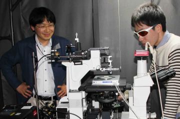
Watching a graduate student set up a Raman microscope
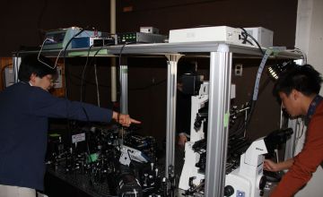
Showing a graduate student how to operate a Raman microscope
One disadvantage of Raman scattering was that it is incredibly faint. Prof. Kano vastly improved this by using a pulse laser as the light source. As the intensity of the directed light (peak power) increases, its interactions with matter also strengthen, greatly increasing the amount of light observed. Furthermore, if a "white" laser that contains various wavelengths is used, a spectrum of all wavelengths can be captured instantaneously without having to change the wavelength and scan multiple times. This technique, called "multicolor nonlinear Raman spectroscopy," is an original invention of Prof. Kano. It is unique in that it measures living tissues (without staining) in three dimensions in a short period of time and shows the spatial distribution of molecules. Another possible dream for the future is to print molecular information with 3D printers.
In recent years, "molecular imaging," which involves labeling of specific molecules with pigments or fluorescent proteins to capture images of their distribution or behavior, has been a popular technique for observing the inside of the human body. Although this technology can visualize biological phenomena in living tissues, only labeled molecules can be seen. In contrast, if Raman spectroscopy is used, it is possible to discern the components of molecules through spectral analysis even when various types of molecules are mixed in together. Prof. Kano's goal is to develop "label-free" molecular imaging that can be used to observe the body in its natural state without labeling. This is because when targets are not predetermined, it is more likely that unexpected things, that is to say, unknown changes and abnormalities, will be discovered.
Prof. Kano's specialty field of research to develop analytical methods and improve the performance of devices would not exist if there were not materials to analyze. In other words, collaboration with people who research those materials is indispensible. That is why Prof. Kano says that the University of Tsukuba is a treasure trove of collaborators. So far, he has collaborated on research about topics such as iPS cells and oil-producing algae. Through interdisciplinary collaboration, he gets to expand the potential for application of Raman spectroscopy while also experiencing the excitement of combining findings with other researchers to make new discoveries. Prof. Kano's motto is "interdisciplinary collaboration on molecular fingerprints."
The important part of performing Raman spectroscopy is the ability to interpret spectra. A vibrational (Raman) spectrum should also be called a "molecular fingerprint." Even if a spectrum is painstakingly measured, the measurement has no significance if the proper information is not taken from it. There are techniques for statistically analyzing spectra, but an expert eye that does not overlook small peaks that resemble noise is necessary to arrive at proper analytical results. This is the first thing that Prof. Kano teaches to students in his laboratory. The outline of Mount Tsukuba viewed from the laboratory looks like a Raman spectrum to Prof. Kano. With this discerning eye, he is searching for the fingerprints of various molecules.
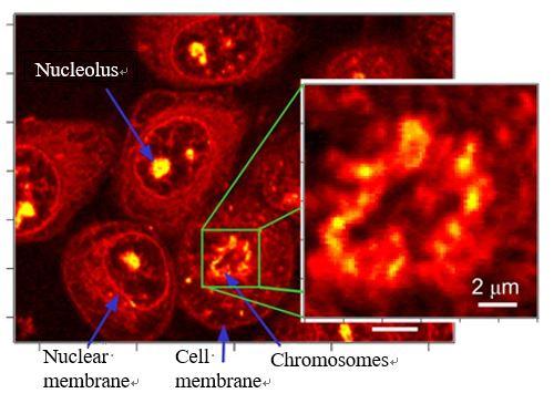
Results of nonlinear Raman imaging of HeLa cells
Article by Science Communicator at the Office of Public Relations