TSUKUBA FUTURE
#061 Measuring with Magnetism
Associate Professor TERADA Yasuhiko, Faculty of Pure and Applied Sciences
MRI (magnetic resonance imaging) devices are used in hospitals for tomographic imaging of body organs such as the brain. Use of such devices involves the patient being put into a tunnel-like gantry and keeping absolutely still until the imaging process is over. Unlike CT scanners, which use X-rays, the MRI scanner uses magnetism so there is no danger of exposure to radiation. In addition to its medical applications, other uses for MRI technology, such as imaging of plants and other living tissues, are being researched.
Every hydrogen atom is essentially like a tiny magnet due to a phenomenon known as nuclear spin. However, when a large number of atoms are clustered together, overall magnetic properties disappear because the directions of each individual nuclear spin are too disparate. Nonetheless, when the cluster is placed in a strong magnetic field, it causes a reaction (resonance), the nuclear spins line up, and magnetic properties are brought back to the fore (NMR; nuclear magnetic resonance). If these signals are measured, tissues with different water content can be distinguished, and this is the principal behind MRI (magnetic resonance imaging) technology. The American and British researchers who developed this were awarded the Nobel Prize for Physiology and Medicine in 2003, in recognition of the fact that this technology had revolutionized medicine, particularly the science of the brain.
Hospital MRI devices are tunnel-like gantries which are big enough to fit completely around a human body. Can these not be made more compact and maneuverable? The idea of using a permanent magnet instead of an electromagnet occurred to the University of Tsukuba Prof. Katsumi Kose, and in 1998 he succeeded in developing the world's first permanent-magnet-based compact MRI device. Since then, improvements have brought about the development of the first mobile MRI device able to produce images of parts of the human body, small animals, plants, living specimens, foodstuffs, materials, fluids, and other objects. Prof. Terada, who was a postdoctoral researcher working in a neighboring laboratory on research and development of a scanning tunneling microscope, was introduced by Prof. Kose to the world of MRI in 2008. Prof. Terada, who had always been interested in manufacturing and measurement, took to Prof. Kose's laboratory like a duck to water.
Nowadays, when research devices are becoming ever bigger and more hi-tech, the MRI is a rare and precious tool in that handmade devices can be used for research. It is true that imaging takes a long time and the images produced are low-resolution, but it enables us to see things which cannot be seen using an optical microscope. Various technologies are used in this MRI, which has a variety of extended applications. A device can be designed, coil rolled, and the system assembled to fit a specific object a researcher wants to take measurements of. Despite the fact that the magnetic field can be adjusted more easily in small devices, these are more difficult to produce. A special device for measuring the bone age of children's hand bones was developed in a joint research project with the University of Tsukuba Hospital orthopedic surgery department, and this is useful for the early diagnosis of dwarfism. X-rays are not recommended for small children because of the dangers of exposure to radiation. However, that said, hospital MRI devices are too large to produce high-quality images of delicate and slender bones. In collaborative research with a plant researcher, attempts were made to measure the growth of rice rhizomes and to monitor the health of fruit trees. Measurement of water conduction in trees enables pear dwarfism to be diagnosed.
Development of the device alone does not solve everything. The results of the measurement carried out in the magnetic field produced by the coil, are also received by the coil. Analysis of this signal waveform data is then output as an image. Occasionally we come across students who are able to master this entire process by themselves, but the norm is for each student to have a specialist field of interest, such as measurement, device construction, or programming. These preferences are taken into account when students are given advice about their research themes. In March 2016, graduate student Akiyoshi Nagata achieved the first-ever visualization of the flow of sap inside a tree growing in natural conditions. By developing a compact MRI that can image trees in situ, he made it possible to image the flow of sap in Japanese zelkova growing outdoors. By continually imaging the flow of sap, he was able to ascertain that sap flow changes considerably between day and night, and before and after leaf drop. However, the role of the physics researcher ends here and further research is now the job of others such as plant physiologists.
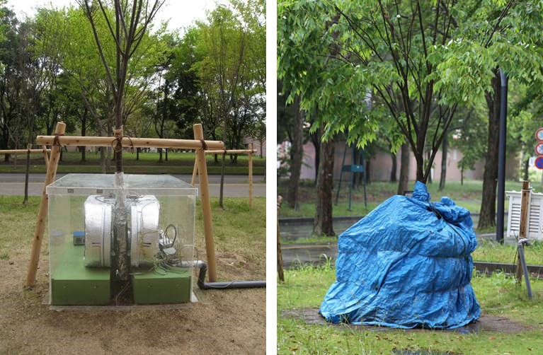
A compact MRI device with a magnetic field of 0.2 tesla attached to either side of a Japanese zelkova tree. The magnet is inside an acrylic box (left), which is then covered with a tarpaulin sheet (right) to protect the device from the wind and rain.
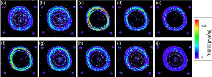
Spectral image cross-sections of the zelkova trunk show the volume flow of sap per pixel (flow volume per unit of time).
A newly assembled device is tested using easily available objects, such as fruit purchased at a supermarket. The team has measured the water content of rice balls and beefsteaks for the popular NHK TV program Tameshite Gatten. The physicist Torahiko Terada (1878-1935), whose exploits reached as far as earthquake research through the X-ray analysis of crystals and who is famous for his observations including that "natural disasters only reoccur once they have been forgotten," wrote his doctoral thesis on the acoustics of the shakuhachi flute, used special methods to photograph bullets in midair, and studied how the angled bulges of konpeito confectionary are formed. This curiosity about everything to do with physics seems to be flowing in the blood of Torahiko's great-grandson Yasuhiko.
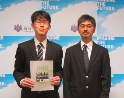
This duo measured sap flow outside in the open air, a spectacular achievement. This photograph was taken at a press conference with graduate student Akiyoshi Nagata (left). He is holding the international academic journal featuring his thesis as the cover photo. (Photograph taken by Prof. Katsumi Kose)
Article by Science Communicator at the Office of Public Relations